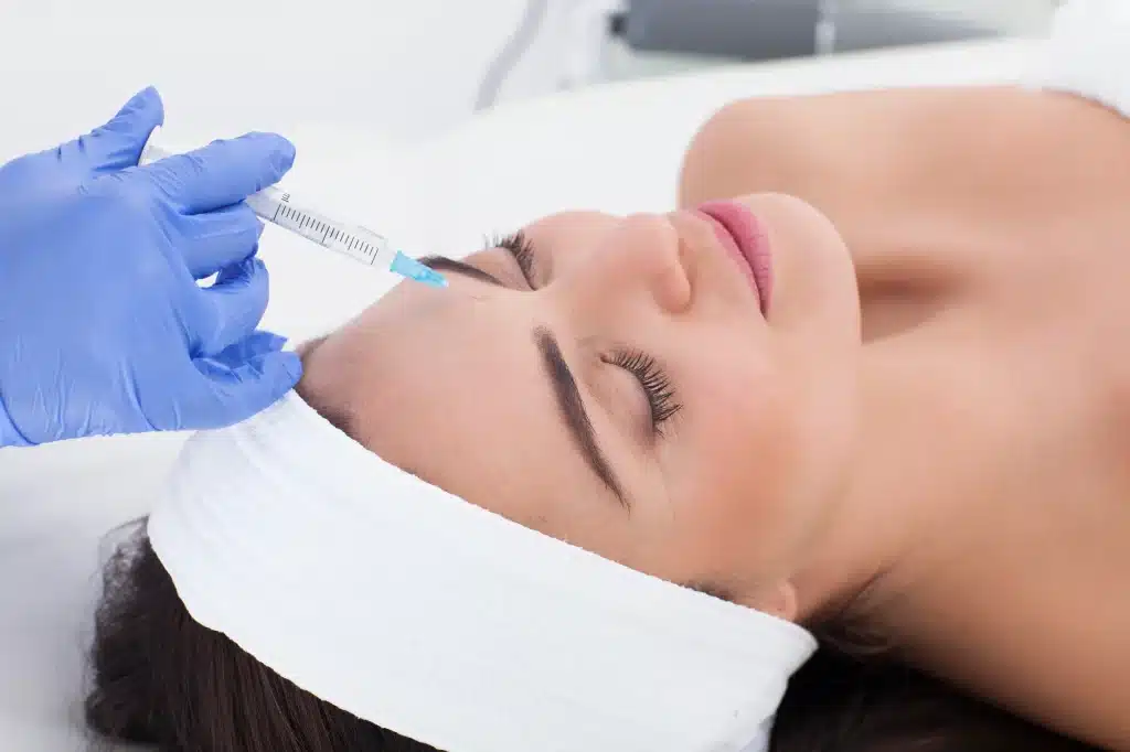
Lip Filler Migration is one of the most frequently encountered aesthetic complications in clinical practice, and one of the most preventable. Although patients often assume migration is caused by product failure, the underlying drivers are usually anatomical or technical. Incorrect depth, volume overload, poor anatomical respect, and repeated stacking without assessing filler persistence are the dominant contributors to displacement.
Migration is not merely a cosmetic issue. It compromises lip mechanics, alters perioral anatomy, and increases the likelihood of fibrosis and long-term asymmetry. In advanced cases, migrated material may disrupt tissue mobility or obscure anatomical landmarks used for safe injecting. The true clinical objective is not simply to restore appearance, but to preserve structural integrity.
This article focuses on how migration happens, how to prevent it, and how to correct it when necessary. Emphasis is placed on injection technique, anatomical restraint, enzymatic management, and long-term control strategies. The goal is not volume correction, but anatomical restoration.
Key takeaways
- Lip filler migration is primarily caused by technique and placement errors.
- The most powerful prevention tool is correct anatomical depth.
- Stacking filler without degradation assessment increases risk exponentially.
- Dissolving migrated filler should be targeted, not aggressive.
- Hyaluronidase must be used selectively and conservatively.
- Migration risk continues after treatment and depends on patient compliance.
- Migration is part of filler safety for injectors and must be managed clinically.


Understanding the mechanics: causes of filler migration
Migration develops in response to mechanical forces acting on poorly anchored filler. The lips are anatomically prone due to high mobility, limited deep fat compartments, and constant compression during speech and mastication. When filler is placed outside stable retention planes, it is gradually displaced toward lower-resistance zones.
Superficial placement is a frequent technical failure. Product positioned near the dermis lacks structural support and is easily moved by swelling and facial motion. Conversely, deep placement without respect for anatomical units creates redistribution pathways that allow product to escape.
Volume stacking is another common driver. Injecting into previously filled tissue without assessing breakdown leads to unpredictable pressure gradients. Over time, filler propagates into the philtrum, vermilion border, or perioral region.
Common causes include:
- Injection outside stable tissue planes
- Excess volume relative to tissue capacity
- Repeated injection without degradation evaluation
- Low-cohesivity fillers in mobile zones
- Poor early swelling control
- Excess lip manipulation after treatment
Edema, bruising, and Tyndall effect often mimic migration in the early phase. True migration evolves over weeks or months and progressively distorts anatomy.
Clinically, migration should be understood as procedural failure rather than adverse reaction. When causative mechanisms are identified early, intervention becomes less invasive and outcomes become recoverable.
Expanded mechanical and anatomical contributors
Lip filler migration is also influenced by tissue quality and vascular dynamics. Patients with reduced dermal thickness, loss of collagen scaffold, or post-inflammatory remodeling exhibit lower resistance to displacement forces. In these cases, filler behaves less like an implant and more like a compressible fluid, redistributing under repetitive motion.
The orbicularis oris plays a central role. This circular sphincter muscle generates multidirectional force during speech, mastication, and facial expression. Filler placed within zones of maximal contraction is subjected to continuous mechanical load, accelerating movement into adjacent low-resistance spaces. Over time, this produces the classic “shelf effect” above the vermilion or irregular expansion into perioral skin.
Anatomical variation must also be considered. The size and contour of the premaxilla, shape of the alveolar ridge, and degree of dental protrusion all influence how soft tissue drapes over bone. Patients with retrusive profiles often receive excessive volume in an attempt to compensate visually, which increases migration risk when structural deficiency is left untreated.
Another frequently overlooked factor is lymphatic and venous drainage. Compromised outflow delays post-injection edema resolution and prolongs tissue pressure, increasing the probability of displacement during the integration window. This is particularly relevant in smokers, patients with autoimmune conditions, or those with chronic inflammation.
Migration is therefore not merely a function of injector error. It is a convergence of tissue mechanics, anatomy, and behavior. Superior technique must account for all three.
Prevention first: how to prevent filler migration
Prevention starts with assessment. Tissue elasticity, prior filler history, and movement patterns determine how injectable material behaves. Ignoring these factors increases risk even when volume is modest.
Depth selection remains the most important technical element. Retention is determined by correct plane selection, not product choice. Filler placed in zones of muscular traction or loose connective tissue is almost guaranteed to migrate.
Another key factor is capacity awareness. The lips cannot be expanded indefinitely. Overfilling may not appear immediately but becomes evident as swelling subsides.
Preventive strategy includes:
- Matching filler rheology to lip mobility
- Conservative volume planning
- Avoiding continuous linear injection across the border
- Depositing filler in support zones
- Staging treatment across sessions
- Assessing old filler before adding new
- Reinforcing anatomical boundaries through technique
Patient education is a preventive tool. Dentition, chewing habits, sleeping posture, and heat exposure influence early filler integration. Movement in the first week destabilizes newly placed product.
Prevention depends less on brand selection and more on anatomical discipline.
Clinical recognition and diagnosis of lip filler migration
Migration rarely presents acutely. It evolves gradually and often asymmetrically. Typical locations include the vermilion border, philtral columns, and perioral skin.
Palpation often identifies mobile or irregular filler beyond intended borders. In chronic cases, fibrosis may be present, making product feel firm.
Diagnostic signs include:
- Loss of clear lip outline
- Product palpable above the vermilion
- Volume inconsistent with injection record
- Distortion of Cupid’s bow
- Asymmetry despite symmetric technique
- Swelling persisting beyond three weeks
Timeline helps differentiate causes. Swelling appears early and resolves. Migration appears late and progresses.
Ultrasound is helpful in complex cases. Imaging may be necessary when chronic accumulation, scarring, or uncertain filler location is suspected.
Migration does not resolve spontaneously. Untreated product may fibrose and complicate future correction. Early detection allows conservative intervention. Late recognition increases complexity.
Active correction: filler migration treatment
Treatment depends on severity, product type, and duration.
Early suspected migration can be observed briefly, but confirmed displacement requires intervention. If structural failure is not corrected, migration recurs.
Treatment options include:
- Observation when diagnosis is uncertain
- Targeted dissolution
- Full enzymatic breakdown when diffuse
- Delayed reconstruction
- Structural re-augmentation
Dissolving migrated filler must be conservative. Blanket enzyme application risks collateral volume loss and contour collapse.
Response differs with filler age. New injections respond predictably. Old filler may resist and require multiple sessions.
Correction must be staged. Enzyme first. Recovery. Reassessment. Rejection attempted too early leads to deformation.
This is not an emergency unless vascular compromise exists. However, delay increases fibrosis risk.
Meticulous documentation supports planning and outcome control.
Decision-making in complex correction cases
Not all migration cases should be treated aggressively. Determining whether to dissolve partially or fully is a strategic choice based on tissue response, patient priorities, and risk profile. In some patients, removing a minimal amount of migrated filler restores balance without disturbing central volume. In others, partial correction fails because residual filler continues to influence soft tissue dynamics.
A systematic approach improves outcomes:
- Assess migration extent clinically and by palpation
- Identify original injection zones
- Map displacement patterns
- Determine whether fibrosis is present
- Decide between focal or global correction
Patients with long-standing filler often exhibit altered tissue architecture. Fibrotic encapsulation restricts enzyme diffusion, which can result in uneven dissolution. These clients require staged correction and extended intervals between interventions. Forcing rapid resolution increases irregularity risk.
Emotional management is also part of treatment. Migration is visually distressing and often associated with loss of confidence. Transparent explanation of process and staged recovery timelines builds trust. Underpromising and overdelivering is preferable to guaranteeing rapid results.
Correction should never be performed impulsively. The strategic injector treats migration as a structural problem requiring sequencing, not a cosmetic defect requiring volume removal.
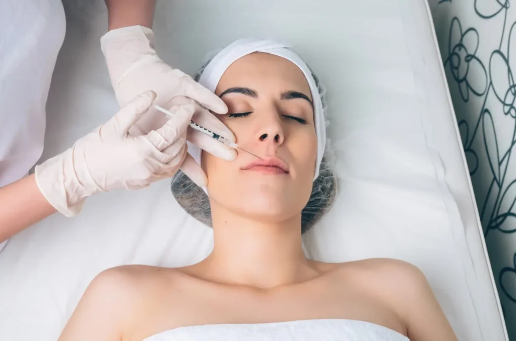
Precision reversal: hyaluronidase correction technique
Enzymatic correction demands restraint. The aim is not eradication but structural restoration.
Mapping improves accuracy. Palpation or ultrasound defines target zones. Initial doses should be conservative and reassessed.
Key principles:
- Inject highest displacement zones first
- Allow time between sessions
- Avoid uniform dosing
- Limit near border
- Document response patterns
Activity evolves over days. Reinjection during enzyme activity enhances distortion.
Complications include erythema, irregular dissolution, and rare allergy. Emergency readiness is mandatory.
Fibrotic tissue often requires staged correction. High cross-linked products dissolve slowly.
Hyaluronidase is also diagnostic. Tissue response informs future placement.
Control and continuity: filler aftercare protocol
The first post-treatment week determines outcome stability.
Lips experience continuous movement. Without modification, even ideal placement may fail.
Aftercare includes:
- No lip massage for one week
- Delay dental procedures
- Avoid heat exposure
- No strenuous exercise 48 hours
- Sleep supine
- No alcohol for 24 hours
Patients must understand early warning signs.
Long-term management favors staged rebuilding in high-risk individuals.
Follow-up enables early intervention. Consistency depends on accuracy, not overcorrection.
Aftercare is structural support, not a formality.

FAQ
Can migration resolve naturally?
No. Swelling resolves. Migration does not.
When can it appear?
Weeks to months after treatment.
Is it inevitable?
No. It increases with stacking and poor depth control.
When is hyaluronidase indicated?
When anatomy is distorted or filler is displaced.
Does enzyme harm natural tissue?
Incorrect dosing may. Conservative technique minimizes risk.
References (AMA)
DeLorenzi C. Aesthetic Surg J. 2014;34:584–600.
Beleznay K, et al. Aesthetic Surg J. 2015;35:623–634.
Pavicic T, et al. J Cosmet Dermatol. 2020;19:2155–2164.
Schelke LW, et al. J Cosmet Dermatol. 2015;14:113–123.
Cotofana S, et al. Plast Reconstr Surg. 2020;145:1159e–67e.
King M, et al. Aesthetic Surg J. 2020;40:NP98–109.
Van Loghem JAJ, et al. J Drugs Dermatol. 2018;17:791–799.
Jones DH, et al. Dermatol Surg. 2020;46:1182–1190.
De Boulle K, et al. Dermatol Surg. 2015;41:S1–S16.
Werschler WP, et al. J Clin Aesthet Dermatol. 2019;12:32–40.
Ghassemi A, Prescher A. Clin Anat. 2012;25:351–357.
Rohrich RJ, et al. Plast Reconstr Surg. 2011;127:240–251.
Dayan SH, Jones DH. Facial Plast Surg Clin N Am. 2019;27:213–232.
Narins RS, Beer K. J Clin Aesthet Dermatol. 2017;10:33–42.