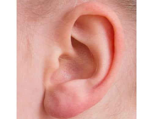
In the cosmetics industry, there has been a recent rise in demand for earlobe restoration and remodeling. Earlobes are prominent features of the overall facial aesthetic, helping to equalize the overall dimensions of the face. As such, large, deformed, or asymmetrical earlobes can throw off facial balances, which may seriously affect one’s self-esteem.
Luckily, various techniques have been identified for earlobe modification; now, there are various techniques with or without dermal fillers, that can help to modify the earlobe to one’s desired fit.
Anatomy
To remodel the ear, each practitioner should be well-informed on both external and internal ear anatomy.
The external ear:
The external ear is the visible part of the ear. This includes the auricle (or pinna) and the ear canal. The auricle is made of tough cartilage and six intrinsic muscles, and consists of the helix, antihelix, lobule, tragus, and concha.
The human earlobe is the soft and fleshy part of the pinna. It consists of areolar and adipose connective tissues, and is less firm than the rest of the auricle. This flap is integral in housing a large blood supply for the ear as a whole, regulating temperature and maintaining balance. The earlobe is unique to each individual and falls into the general category of “attached” or “detached,” in which the lower lobe is either connected to the anterior inferior side of the cheek or hangs freely.
The internal ear:
The internal ear is comprised of a variety of nerves and arteries.
- The blood supply systemAn extensive network supplies blood to various parts of the ear. The main external carotid artery consists of three branches, which are the occipital artery, posterior auricular artery, and the anterior auricular branch of superficial temporal artery.
- The nervous systemThe four main sensory nerves that innervate the ear are:
- The great auricular nerve, which is connected to the lower 2/3 of the anterior and posterior external ear, including the earlobe;
- The auriculotemporal nerve, which is connected to the anterior superior 1/3 of the ear including the tragus, crus, helix and superior helix;
- The lesser occipital nerve, which innervates the posterior surface of the superior 1/3 of the auricle;
- And the auricular branch of vagus nerve, which innervates the external auditory canal floor and concha.
Treating earlobe concerns
The most common cause of trauma and damage to the ear is ear piercings. Some earrings may cause irritation or injuries such as split earlobes, enlarged ear piercing holes, and keloid scarring.
Split earlobes
Individuals who wear large and heavy earrings are at risk of experiencing a cut or a split through the earlobe. Minor occurrences, such as accidental pulls from the hair or movement in sleep may cause this injury. Therefore, it is advisable for wearers to take their earrings off before going to bed.
Enlarged ear-piercing hole
Enlarged plugs, or “gauges,” are a common choice of fashion for the ears among young patients. Believed to be partly originating from the body art movement throughout the 1980s, this method or decorating the ear involves wearing plugs or gauge earrings that are frequently changed to gradually stretch and expand the opening in size, starting at 1mm to more than 2cm large. It leaves a large gaping hole in the earlobe, the desired result of these patients.
Should it be desired, to reduce the size of this hole, simply reverse the process with the size of the earrings. However, this does not guarantee a complete reduction in size, especially when one has an overly stretched hole. Relatedly, due to the increase of gauge wearers over the years, the demand for earlobe repair in clinics has become higher than ever.
Guide to treatments
It is important to take note that there is no one-size-fits-all approach in treating these cases: each case requires a specific technique in order to achieve the best possible result. The preferred treatment techniques are outlined below.
Split earlobe
When it comes to repairing split earlobes, the patient and the practitioner have the choice to either reconstruct or not reconstruct the piercing’s hole. Certain doctors prefer to dismiss the reconstruction method and advise patients to wait for at least 6 months before re-piercing the ear, situating it at 2mm medial or lateral to the previous scar.
For earlobe repair, the practitioner can create a straight line or a broken line of repair. Although the straight-line repair method requires minimal effort, its final result may not be as aesthetically pleasing as the broken-line method, as its scar will eventually contract and form a notch at the free border of the ear.
Other alternatives to the straight-line technique may include Z-plasty, the lab-joint technique, or L-plasty. Z-plasty is a technique that forms a Z-shape wound. This wound aims to prevent intense scarring of the earlobe.
L-plasty is highly recommended for incomplete split earlobe repair. Cutting the skin around the edge of the split and stitching it back together may leave an extension of the earlobe, which may not be desired by patients.
Earlobe reduction
In earlobe reduction and remodeling, the earlobe tissue should be removed without leaving any prominent scarring. The following method, found by Dr Salinda Johnson, has a high success rate on reducing the possibility of notch formation.
Before beginning any procedure, the practitioner should consult with the patient on their desired lobe shape. They should draw the desired shape onto the ear using a white marker. Then, they should draw one line from the tragus to the white mark level, and a second line on the earlobe from the tragus to the lateral end of the white mark. After measuring the length of both lines, they must create a triangle, ensuring that the size of the triangle base is similar to the length of the first and second line. Then, they should carefully excise the earlobe as marked. The first and second lines should then be joined using deep dermal and vertical mattress stitches.
Approaching the procedure
Because the ear is highly innervated, it is important to carry out earlobe repair under local anesthesia to reduce the amount of pain. Carry out the treatment as follows.
- For L-plasty, draw the lines of excision on the highest point of the cleft before the edges curve gently. If these lines are not accurate, a groove will be left along the repair line, giving the impression of a notched border.
- Inject 2% xylocaine with epinephrine or 2% lidocaine at the junction of the earlobe and cheek to avoid the risk of facial nerve injury. Alternatively, you can infiltrate the anesthetic solution directly into the earlobe until it becomes firm and pale for easier scalpel excision.
- For L-plasty and Y-plasty techniques, excise the areas from the free border to the apex of the cleft of the earlobe using an iris scissor or scalpel. Check that the backside of the earlobe has been cut to the same shape as the front. Then, insert the flap accurately beginning from the anterior side. Ensure there is no irregularity in the free border of the earlobe.
- Use non-absorbable sutures to stitch the desired shape with a vertical mattress technique. This method prevents the scar from rolling inward as it heals.
Avoiding complications
- Cut through the earlobe properly by checking the backside of the earlobe before stitching the wound.
- All stitches should be tightened properly to avoid any post-procedure bleeding or accidental wound openings.
- Stress or applied pressure on the area may loosen up the stitches. Patients should sleep on their backs and avoid playing contact sports to protect the ear.
- With precision, ensure that both sides of the ears have a similar shape by measuring the markings made on both earlobes. Ask the patient to sit up straight to check if they have the same angle and length. Prior to the procedure, inform the patient that their earlobes may not be symmetrical, as results are largely dependent on the available tissue.
- If the stitches are not tight, bleeding, bruising, and hematoma may occur. Patients should avoid taking blood-thinning substances such as alcohol, ibuprofen, aspirin, and vitamin C and E prior to these procedures.
- In treating ear keloids after surgical excision, triamcinolone (TCN) may cause an anaphylactic reaction or type 1 anaphylactic reaction with immunoglobulin E (IgE antibodies). Prior to treatment, investigate if the patient has a history of allergic reaction to steroids.
- Patients with skin types V-VI may experience scarring. Patients who have a history of keloid or hypertrophic scars should be advised that keloid scarring could occur.
- Advise patients to avoid getting the treated area wet for the first 2 to 3 days after surgery.
A variety of earlobe repair treatments are available for various conditions. Doctors should be aware of choosing the right technique in order to achieve satisfactory results.

